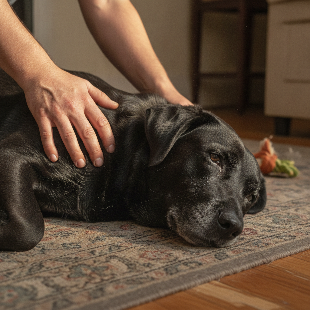
Your hand runs over your dog’s back, the way it has ten thousand times before, and stops. There’s something there. Something that was not there last week.
What happens next is almost always the same. You press it. It’s soft. You press it again. It moves a little. You tell yourself it’s probably a fatty thing, because a friend’s dog had a fatty thing, and you decide to keep an eye on it.
Here is the single most useful sentence in this entire article: you cannot tell what a lump is by touching it, and neither can your vet. Not by feel, not by looks, not by size. A malignant mass and a harmless one can be indistinguishable to the fingertips. The only thing that answers the question is a needle and a microscope.
Which means “keep an eye on it” is not a plan. It’s a delay. And delay is the one thing that reliably makes a bad outcome worse.
The rule that tells you when to stop watching
Veterinary oncology has a memorable shorthand for this: See Something, Do Something. Why Wait? Aspirate.
The working guideline is straightforward. If a skin or under-the-skin mass is at least the size of a pea (about 1 cm) and it has been there a month or longer, it should be sampled, not monitored.
The test is called a fine needle aspirate. It’s fast, it usually costs less than people expect, it typically requires no sedation, and it’s done in a normal appointment. A thin needle collects a few cells, they go on a slide, and someone looks at them.
The reason this rule exists is that the cost of aspirating a lump that turns out to be nothing is roughly one appointment. The cost of watching a mass cell tumor for eight months is a much bigger surgery, sometimes a much worse prognosis, and sometimes no good options at all. The asymmetry is brutal, and it’s the whole argument.
“I have never once regretted aspirating a lump that turned out to be fat,” says Dr. Mara Chen. “I have absolutely regretted watching one. The needle is cheap. Time is not.”
What you actually can learn by feel
Not the diagnosis. But observation still helps your vet, so make it good. Note:
Is it on the skin, in the skin, or under it? If you feel a bump under the surface with no disruption to the fur or the outer layer of skin, that points one direction. If there’s missing fur, scabbing, or redness on the surface, the problem is likelier to be with the skin itself. That distinction genuinely narrows the list.
How fast is it changing? A mass that’s the same size in six months behaves differently from one that doubled in three weeks. Take a photo next to a coin, with the date. Do it monthly.
Is it fixed or does it move? A mass that slides freely under the skin behaves differently from one that feels anchored to the tissue beneath it.
Is it bothering your dog? Licking, chewing, or flinching at the spot is information.
Bring all of that. But bring it as observations, not as a conclusion.
The common causes, and why none of them can be diagnosed by eye
Lipomas. Benign fatty tumors, common in middle-aged and older dogs. They’re always under the skin, they feel soft and squishy, and they usually need no treatment at all if they’re small and slow-growing. Some do get removed, either because they grow fast or because they sit somewhere that makes them uncomfortable, like an armpit or behind a knee. The catch, and this is the whole point: other tumors, including malignant ones, feel and look like lipomas. A diagnosis of lipoma is a diagnosis a vet makes with a needle, not a guess you make with a thumb.
Skin tags. A catch-all for growths that dangle off the skin’s surface. The medical terms cover a range of different things, including sebaceous gland adenomas, acrochordons, sebaceous cysts, apocrine cysts, and follicular cysts, each with its own characteristics. Malignant growths can mimic all of them, so a biopsy is what settles it.
Pigmented growths. Anything black, bluish, or darkly red-tinged. If it has color in it, biopsy it. Plenty of pigmented masses turn out to be harmless. Others turn out to be melanoma, and there is no way to sort them by eye.
Hives. Round, flat or domed swellings from an allergic reaction, sometimes itchy and sometimes not. Triggered by contact with a plant or chemical, or by something ingested, including food and medication. Hives need veterinary treatment right away, because the underlying allergic reaction can escalate.
Ticks, and flea damage. An embedded tick genuinely feels like a small growth on the skin, and people find them by touch all the time. Fleas are too small to feel, but a heavy infestation causes irritation, which causes bumps and scabs across the dog.
Insect bites and stings. Usually resolve on their own without you ever noticing. But some dogs react allergically, with significant swelling around the site, and that needs a vet the same day.
Skin infections. These cluster in dogs who already have allergies or another underlying skin problem. Anything that breaks down the skin’s normal barrier opens the door to bacteria or yeast. Look for itch, a bad odor, redness, bumps, or scabs. These need treatment for the infection, plus ongoing management of whatever underlying condition is causing them.
Tumors, benign and malignant. Dogs get many kinds. Fine needle aspirates and biopsies are how you tell them apart. Treatment ranges from monitoring, to surgical removal, to chemotherapy or radiation, and it depends entirely on the type, which is another way of saying it depends entirely on the sample.
And the odd ones. Older dogs with Cushing’s disease can develop mineralized bumps called calcinosis cutis, plus comedones (blackheads). Dogs with autoimmune disease can develop vasculitis, causing scabbing in specific areas. Traumatic injuries leave scars and, from repeated microtrauma, skin tags. Viral infections cause warts, or papillomas.
The list goes on long enough that visual identification is not a skill anyone has. Not you, and not a vet with thirty years of experience. That’s not a knock on vets. It’s how skin works.
What to actually do this week
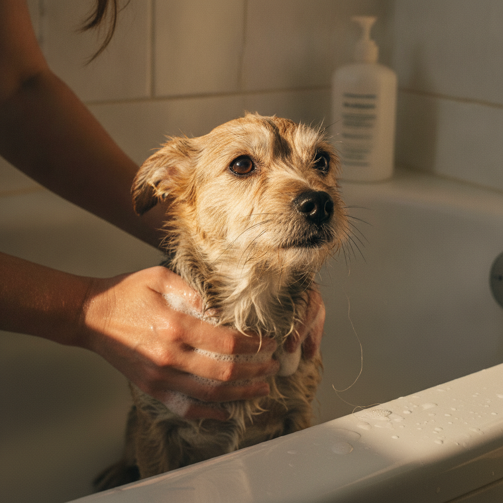
- Find them all. Once a month, run both hands slowly over your entire dog: back, chest, belly, legs, armpits, groin, neck, under the collar, around the tail. Bumps are much easier to feel when a dog is wet, or right after a groom, so bath time is a good time.
- Map them. Keep a simple lump map: a rough drawing or a note with locations, sizes, and dates. Photograph each with a coin for scale. When a mass changes, you’ll know, instead of arguing with your own memory.
- Apply the rule. Pea-sized or bigger, and there a month or more? Book the aspirate. Growing fast, changing color, hard, fixed in place, ulcerated, or bothering your dog? Book it now, regardless of size.
- Recheck the old ones. A mass diagnosed as a lipoma two years ago that suddenly feels different is a new mass as far as your vet is concerned. Old diagnoses have a shelf life.
What newer research adds
The direction of veterinary oncology over the last decade has been consistently toward earlier sampling, and the reason is simple: early-detected skin and subcutaneous tumors are dramatically more treatable, and the diagnostic step is cheap and low-risk. That’s the entire logic behind the “why wait, aspirate” campaigns you’ll increasingly hear from vets, and it’s why the phrase “let’s just monitor it” is much rarer in a good clinic now than it was ten years ago.
The other useful update is on lipomas specifically. Large primary-care studies in the UK confirm that lipomas are extremely common, are overwhelmingly managed conservatively, and rarely need to come out. That’s reassuring, and it is also exactly why the aspirate matters: the whole value of confirming a lipoma is that it lets you confidently stop worrying about it. You can’t buy that peace of mind with your fingertips.
References
- Ettinger, Sue, and Erin Bannink. “Veterinary Oncology: What to Do With Lumps and Bumps on Dogs and Cats.” Today’s Veterinary Practice. https://todaysveterinarypractice.com/oncology/tumor-detection-dogs-cats/
- Pegram, Camilla, et al. “Clinical Management of Lipomas in Dogs Under Primary Veterinary Care in the UK.” Veterinary Record, vol. 187, 2020. https://doi.org/10.1136/vr.105804
- Heinrich, Nicole A. “Urticaria (Hives, Wheals) in Animals.” Merck Veterinary Manual, 2024. https://www.merckvetmanual.com/integumentary-system/urticaria/urticaria-hives-wheals-in-animals
- American Animal Hospital Association. “AAHA Oncology Guidelines for Dogs and Cats.” https://www.aaha.org/resources/2026-aaha-oncology-guidelines-for-dogs-and-cats/
- American Veterinary Medical Association. “Cancer in Pets.” https://www.avma.org/resources/pet-owners/petcare/cancer-pets
- Brooks, Wendy. “Benign Sebaceous Gland Tumors.” Veterinary Partner, VIN. https://veterinarypartner.vin.com/default.aspx?pid=19239&id=4952215