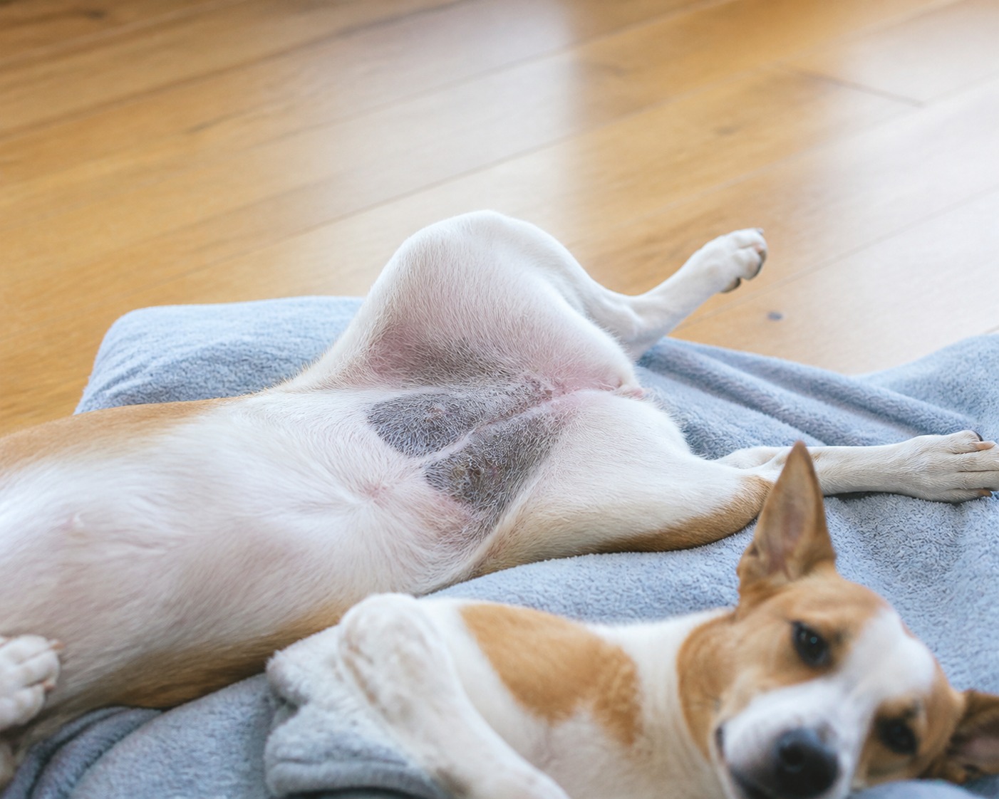
You are scratching your dog’s belly and you notice it: a patch of skin that used to be pink has gone gray, or almost black. It can be alarming, and the instinct is to treat the color itself as the disease. That is the wrong way to look at it. Hyperpigmentation is not really a condition you catch. It is your dog’s skin keeping a record. The darkening is the paper trail of something else, and reading it correctly is the whole game.
Here is the reframe worth holding onto: in the vast majority of cases, the dark skin is a symptom, most often the aftermath of inflammation. Chase the color and you will get nowhere. Find what has been irritating, itching, or hormonally disrupting the skin, treat that, and the color usually follows the cause back to normal.
What you are actually looking at
Hyperpigmentation simply means skin, or sometimes the fur itself, has increased its pigment and turned darker, typically to gray or black. It tends to appear where fur is naturally sparse, which is why owners spot it first on the belly, in the armpits, or around the groin.
The color change rarely travels alone. Depending on what is driving it, the same patch may also show:
- Discoloration, the darkening of skin or fur in focal areas.
- A change in texture, with skin becoming thickened, dry, or crusty.
- Hair loss, so the darkened area also looks thin or bald.
- Odor, especially when a skin infection is part of the picture.
- Itchiness, with a dog scratching or licking the spot.
- Whole-body signs when the cause is systemic, such as unexplained weight gain, low energy, and a noticeable jump in how much your dog drinks and pees.
That last group matters. A dark patch plus a dog that is suddenly drinking and peeing more is a very different conversation than a dark patch on its own, and it points the investigation somewhere specific.
The one exception: skin that darkens on its own
Almost all hyperpigmentation is secondary, meaning something else caused it. There is one notable exception, and it belongs to a single breed. Dachshunds can develop a primary form, called acanthosis nigricans, where the skin, usually in the armpits and groin, darkens without any other underlying trigger. It is genetic, particular to the breed, and not the result of an infection or illness. If you have a Dachshund with darkening in those folds, this is the possibility your vet will weigh.
Why most dark patches show up: the causes
For every other dog, hyperpigmentation is the skin’s response to some ongoing insult. The common drivers:
Chronic inflammation. This is the headline cause. Anything that keeps skin inflamed over time can leave darkening behind. Most of the causes below are really just specific routes to the same inflamed state.
Skin allergies. Allergies and the allergic skin condition known as atopy keep skin itchy and inflamed. When that is not well controlled, discoloration can follow.
Contact dermatitis. When something touching the skin directly irritates it, grasses, plants, dust, or other materials, the resulting inflammation can eventually darken the area.
Skin infections. Bacterial, yeast, and fungal infections, along with fleas and skin mites, all generate heavy inflammation that can lead to hyperpigmentation.
Hormonal imbalances. Systemic diseases such as Cushing’s disease and hypothyroidism change the skin, and darkening can be one of the visible clues.
Friction. Skin that is constantly rubbed gets inflamed and can darken. This shows up where a collar or harness chafes, and in overweight dogs whose skin folds rub against each other.
“I tell owners to think of the dark patch as a smoke alarm, not the fire,” says Dr. Priya Nair. “It is genuinely useful information. It is telling you the skin has been under stress in that spot for a while. Our job is to trace it back to the source, because bleaching creams and home remedies aimed at the color itself completely miss the point.”
Some breeds are more prone to it
Any dog can develop hyperpigmentation, but some breeds are predisposed, largely because they are also prone to the underlying skin problems that trigger it. Breeds that show up more often include Dachshunds, Yorkshire Terriers, Australian Shepherds, Pugs, Bulldogs, French Bulldogs, Staffordshire Terriers, Boston Terriers, Shar Peis, Cocker Spaniels, Shih Tzus, and Poodles. If your dog is on that list, it is worth being a little quicker to investigate new dark patches.
How a vet works out the cause
Because the darkening is visible, the diagnosis of hyperpigmentation itself is straightforward, your vet can see it during an exam. The real work is figuring out why, using your account of what you have noticed at home alongside the dog’s other signs, such as itching or systemic changes. To pin down the cause, a vet may reach for:
Skin tests. Impression smears, tape cytology, and skin scrapes look for parasites and bacterial, fungal, or yeast overgrowth.
Allergy testing. For a chronically itchy dog with allergy signs, testing helps. Worth knowing: intradermal skin testing is the method vets trust, while allergy blood panels are unreliable enough that most vets do not lean on them.
Blood and urine testing. Routine labs screen for the systemic changes that come with infection or a hormonal disorder.
Response to treatment. Sometimes the answer confirms itself. If clearing up a skin infection also clears the discoloration, that pins down the cause after the fact.
Treating it means treating the cause
The most effective treatment is to identify and target whatever set off the skin. Once your vet knows the driver, the plan usually combines a few approaches:
Treat the underlying condition. If there is a specific culprit, an infection, allergy, hypothyroidism, or Cushing’s, follow the prescribed plan, whether that is a topical, an oral medication, or both. Ask your vet whether it is a short course or a long-term therapy, and when to come back for a recheck.
Bring down the inflammation. Since darkening is a response to inflammation, reducing it helps the skin recover. The best lever is treating the exact cause, but medicated soothing baths and omega-3 fatty acid supplements can further calm irritated skin. Keeping your dog on year-round flea prevention protects against the intensely irritating bites that inflame skin in the first place.
Remove friction. Where materials rub, reduce the rubbing. If a collar or harness sits over a darkening spot, switch styles for a while and remove it any time it is not actively in use. For an overweight dog, a vet-guided weight-loss plan eases the chafing that comes from deep skin folds.
The takeaway
Darkened skin is a marker of inflammation, not a disease in its own right. It has many possible causes, from infections and allergies to hormonal conditions, and the reassuring part is that it can usually be reversed once the real cause is found and treated. The move is not to panic about the color. It is to notice it early, note anything that comes with it, and let your vet trace it home.
What newer research adds
The core understanding of hyperpigmentation as a marker of underlying skin disease has held steady, and current veterinary references, including the Merck Veterinary Manual’s guidance on primary acanthosis nigricans and postinflammatory hyperpigmentation, continue to frame it that way. What has improved most for owners is the toolkit for the common underlying cause: allergic skin disease. Newer targeted therapies now let vets control chronic itch and inflammation more precisely than older broad steroids did, which means the inflammation that drives darkening can often be shut down sooner, and with fewer side effects.
The takeaway is encouraging. If your dog’s dark patches trace back to allergies, the odds of getting both the itch and the discoloration under lasting control are better now than they were a few years ago. It still starts the same way, though, with a vet visit to name the cause.
References
- Bajwa, Jangi. “Cutaneous hyperpigmentation in dogs.” The Canadian Veterinary Journal, vol. 63, no. 1, January 2022, pp. 85-88.
- Moriello, Karen. “Primary Acanthosis Nigricans and Postinflammatory Hyperpigmentation in Dogs.” Merck Veterinary Manual, January 2025.
- Kim, Ha-jung, et al. “Melanotrichia in Poodles: Clinical and Histopathological Findings.” Journal of Veterinary Clinics, vol. 22, no. 2, 2005, pp. 153-156.