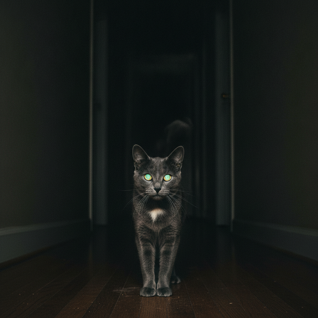
You walk into a dark room and two discs are floating there, pointed at you. It is the single most reliable way a cat can make the hair go up on the back of a mammal’s neck, and it has been doing so for a very long time. Our ancestors saw those discs in the dark and correctly concluded that something was watching them.
But here is the thing nobody tells you about the glow: it is information, and it is free.
The color of your cat’s eyeshine, its brightness, and whether the two eyes agree with each other are all readouts of the anatomy behind those eyes. Vets look at this. You can look at it too, in your hallway, with your phone. Once a month is plenty. And you should know what a bad reading looks like, because the eyeshine can change before anything else about your cat does.
So: first what the mirror is, then how to read it.
They do not glow. Nothing about a cat glows.
“Glowing” would mean producing light. Cats have not developed firefly genes, which is a shame for everyone.
What they have is a tapetum lucidum: a reflective sheet sitting behind the retina, between the retinal epithelium (where the light-detecting cells live) and the choroid (the layer supplying blood to the retina). Light enters the eye, passes through the retina, and any photon that misses a rod or a cone would ordinarily be absorbed and wasted. The tapetum bounces it straight back through the retina, giving the photoreceptors a second chance at it.
Some of that returning light escapes back out through the pupil. That is what you are seeing. You are looking at your cat’s leftover photons.
And the structure doing it is stranger than you would guess. A cat’s tapetum is a highly organized crystal, roughly 15 to 20 cells thick at the center and thinning toward the edges. The cells are packed with tiny rodlets of riboflavin, vitamin B2, arranged in a hexagonal lattice and bundled together. Your cat’s night vision is, in a real and literal sense, built out of a vitamin.
Why the eye was built this way
The tapetum is the last and most dramatic feature in a whole chain of adaptations for low light:
- A large domed cornea. Compare how much of your cat’s eye is clear window versus how much of yours is. Far more of a cat’s eye is dedicated to letting light in.
- Wide pupils. Much larger than ours at full dilation, delivering more of the available light to the back of the eye.
- A rod-heavy retina. Rods fire at low light levels; cones give you color and sharpness. Cats carry a far higher proportion of rods than we do.
- A very reflective tapetum. More reflective than a dog’s, which is a large part of why cats out-see dogs in the dark.
Stack those and a cat needs only about one-sixth of the light a human requires to see. Cats are crepuscular, most active at dawn and dusk, and every one of these features is aimed at that window.
None of it was free. Cats bought low-light sensitivity with sharpness. A cat’s visual acuity is estimated at around 20/150, meaning your cat has to be more than seven times closer to an object than a person with 20/20 vision in order to resolve the same detail. They are superb at detecting a movement in the dark and mediocre at working out what it was. Reading is entirely off the table.
“The tapetum is the most visible part of a trade-off that happened millions of years ago,” says Dr. Ravi Mehta. “Your cat gave up detail to own the dark. The glow is just the receipt.”
Now the part you can actually use: reading the shine
Here is where this stops being trivia.
Normal adult eyeshine is green or yellow. That is the most common reflection you will see, and it is the tapetum doing its job.
Kittens often start blue. A bluish reflection in a young kitten is expected, and it typically shifts to the adult green or yellow around three to four months of age as the tapetum matures. A slow, gradual change in a kitten is not a problem.
Red usually means no tapetum. It is uncommon, but the mirror does not always show up. A few cats simply never develop one, and others end up with a thin or patchy version. With no mirror behind the retina, the light bounces instead off the blood-rich tissue back there and comes home red, which is the same effect as red-eye in a flash photo of a person. These cats likely have somewhat worse night vision than other cats. They are also, thanks to every other feature on the list above, still running rings around you in a dark hallway.
Small differences between the two eyes can be normal, because the tapetum does not always develop identically on both sides.
And here is the line that matters:
A sudden change in the color, brightness, or symmetry of an adult cat’s eyeshine is a reason to call your vet, promptly.
A reflection that abruptly changes color, dulls, brightens, or goes badly asymmetric can indicate a problem with the retina itself, including detachment or bleeding, or inflammation inside the eye. The tapetum sits directly behind the retina; anything that disturbs the retina disturbs the reflection. That is why the glow is diagnostic. You are looking at a structure that is very hard to see any other way.
How to actually check it
This takes about a minute and you already own the equipment.
- Take a flash photo of your cat in a dim room, straight on, at their eye level. Eyeshine is easy to capture; a phone flash does it reliably, and even a non-flash low-light photo will often catch it because those eyes take in so much light.
- Look at the two eyes side by side. Same color? Similar brightness? Same shape of reflection?
- Keep the photo. That is the entire point. A single reading tells you nothing. A photo from six months ago, next to one from today, tells you whether something changed.
- If something changed, do not wait. Retinal detachment in cats is frequently caused by high blood pressure, often secondary to kidney disease or hyperthyroidism, and it can take a cat’s sight in days. Caught early, the underlying cause is treatable and vision can sometimes be saved. Caught late, it usually cannot.
That last paragraph is the real reason to bother with any of this.
What’s new since this was written
Two developments are worth knowing.
The first is that the tapetum has become genuinely interesting to physicists. Work across a range of species has shown that these reflective layers behave as multilayer photonic structures, sub-wavelength gratings whose spacing tunes exactly which wavelengths get bounced back, rather than acting as a simple mirror. That is why eyeshine color varies so much between species and individuals: it is an optical property of the lattice, not a pigment. Your cat’s eyes are running interference optics.
The second is more practical. Veterinary medicine has moved firmly toward screening senior cats for hypertension, precisely because the eye is often where high blood pressure shows up first and where the damage is permanent. Blood pressure measurement is now a routine part of good senior cat care, and sudden blindness or a retinal bleed is one of the classic presentations that brings an undiagnosed hypertensive cat through the door.
So the folk version of this topic ends at “cool, spooky, evolution.” The useful version ends here: look at the glow, photograph it occasionally, and treat a change in it as an early warning rather than a curiosity.
References
- Ollivier, F. J., Samuelson, D. A., Brooks, D. E., et al. (2004). Comparative morphology of the tapetum lucidum (among selected species). Veterinary Ophthalmology.
- Cornell Feline Health Center. Feline Vision: A Cat’s Eye View.
- Taylor, S. S., et al. (2017). ISFM Consensus Guidelines on the Diagnosis and Management of Hypertension in Cats. Journal of Feline Medicine and Surgery.
- Maggs, D. J., Miller, P. E., & Ofri, R. Slatter’s Fundamentals of Veterinary Ophthalmology — retina and tapetum lucidum.